In an isolated bunker beneath a Jerusalem hospital lies a particle accelerator, called a cyclotron • Dror Feuer entered the inner sanctum to try and understand how state-of-the-art radioactive materials are produced here — materials that, after being injected into cancer patients, act as guided missiles to search for and destroy targets • Special for G.

The soundtrack, four floors underground in the basement of the nuclear medicine department at Hadassah Ein Karem Hospital, is one I know from thousands of hours of sci-fi shows and movies. It’s like being on the bridge of the Starship Enterprise. No noise from the world above makes it down here; all you hear is mechanical beeps and digital rings — blip-blip, gling-gling, and of course, toot-toot, eee-eee, trrrr, and above all a thin layer of continuous hissing.
Here, between 2.5-meter-thick walls, with cooling and air systems sealed off from the rest of the hospital, reside two particle accelerators — cyclotrons. One is old, about twenty years, and one newer, about seven years old. They look the same. Each has its own bunker, its own control and monitoring room, cooling room, maintenance, and electronics. On the way there, we pass through these spaces, and I’m thoroughly enjoying the charming contrast between high-tech and a delightful mess: piles of scattered papers, crates, devices, bottles, and a cozy coffee corner leading to a maintenance room filled with saws, hammers, screwdrivers, and tools I did not expect in a particle accelerator. Very Israeli. The control rooms are filled with dials and instruments, some running forward, some backward.
We enter the inner sanctum. The accelerator dominates most of a small room, with thousands of colorful tubes entering and exiting. It looks like the forbidden lovechild of a spaceship and a pressure cooker. Just over $2 million apiece. It even looks a bit alive. I can’t decide if I expected more or less — but either way, I’m happy. Every time I touch something, someone yells at me: “Don’t touch!” Then someone comes through with the Geiger counter, which beeps happily. All is well, they reassure, nothing dangerous. So, every time they’re not looking, I touch.

Inside the accelerator is a vacuum, like space, created by four diffusion pumps reaching a pressure of 10 to the power of minus 7 atmospheres. So, explain to me what’s happening here, I ask Prof. Eyal Mishani, head of the cyclotron unit. “To create a nuclear reaction,” he replies, “we take charged particles, accelerate them in electric and magnetic fields, and then bombard other, stable materials.”
Sure. Now, slower, please.
“First, you create a charged particle. In our case, you take hydrogen gas, feed it inside, and put it through a high electric field. This breaks it up and creates plasma. Plasma is chaos. Out of this, with another electric field, I extract a different particle that, because it has mass and electrical charge, begins to spiral in the electric and magnetic field, gaining speed and energy each revolution until it reaches 18 mega electron-volts. Then, I shoot it out of the field into the target, where it’s bombarded with high-energy protons, creating a new element called F18 – that’s the radioactivity.”
“Picture that, inside this crazy vacuum, there’s no collisions, so I can focus a beam to millimeters,” Mishani continues. “We cool the whole process with helium to minus 180 degrees Celsius. What comes out — a radioactive solution — I send through thin pipes to the lab, where the radioactivity is first separated from the water and then loaded onto the smart molecule.”
“This Is True for Many Diseases”
Mishani, 54, notices that while my body made it down here, my mind hasn’t caught up.
“Let’s say you have a military hangar in Syria,” he illustrates. “You send a plane to photograph it from above and now you know everything about its configuration, location, shape, fences, etc. But you have no idea what’s inside — tanks, planes, rockets. That’s basically anatomical imaging, like X-ray, CT, ultrasound, or MRI. But that’s not enough, right?”
Of course not.
“The world is moving towards personalized, targeted medicine, which demands much more complex and clever solutions. For that, you need information. How do you get it? There’s an invasive way — surgery, biopsy, pathology — which is great, but has issues: it takes time, it’s painful, and samples must be taken from different regions. Then, there’s another way — molecular imaging.”
“If I continue the analogy,” he says, “instead of sending a plane, I send in special elite unit who go inside and report exactly what’s happening. That’s molecular imaging — it lets you see things at the cellular level. How? We inject a radioactive material — a smart compound. A guided missile. It enters the bloodstream and targets specific elements of the tumor. These materials we manufacture here, in the accelerator. I’m essentially launching into a part or tissue of the human body a spyware, which constantly sends back signals. The smart material knows where to go, the radioactive isotope knows what to do when it gets there.”

Unlike in anatomical imaging, with such type of imaging (PET-CT) you can diagnose, with high accuracy, for instance, the presence of a unique type of lung cancer, which is found in only 20% of lung cancer patients. So instead of blitzing the whole body, you can load radioactive isotopes onto smart compounds, which can be injected to the bloodstream. “The isotope sends us signals, and as it accumulates in the vicinity of the tumor, we know whether the molecular element we wish to hit/treat is there or not,” Mishani says. “And this works for many diseases. For example, understanding brain processes, like in Alzheimer’s disease, where beta-amyloid plaques accumulate. There are compounds capable of finding these plaques long before symptoms start. It’s very good for early diagnosis, even if there is no cure yet – and we manufacture these compounds here.”
If you’ve found a tumor, why just send signals and not blow it up?
“If there are neuroendocrine tumors (see glossary), we treat with a compound that reaches and destroys them. That’s the direction, but it’s important — it won’t replace biopsies nor other treatments.”
And in the future?
“As with everything, it will be a combination of tools — one of modern medicine’s tools, for more efficient targeting and monitoring treatments. Another advantage of this imaging – it allows earlier monitoring of treatment efficacy that CT or ultrasound do. If you administer a biological drug to kill a tumor, with CT you’ll see a result only when the mass disappears – that may take four months. Here, you can see it immediately. But it’s complex, difficult, expensive, and not simple.”

How about dangerous?
“We administer the compounds in minute masses. Actually, these materials have no mass, only an electric charge. So, no toxicity problems. The radiation is very specific and we know how to read it. These are unique materials; many don’t last long — they have a short half-life. Some last two minutes or less; the longest half-life we have here is 110 minutes. That means you can’t import or store it — you must manufacture on a daily basis.”
The Difference Between a Reactor and an Accelerator
We stand in front of the accelerator, running now at 515 milli-amps, with internal pressure at 31 bar, and protons bombarding at 75 micro-amps. “Today, we produced 5.5 Curie, which is a lot,” says Mishani. He produces radioactive materials that are similar to glucose and highly accumulate in the brain, bladder, and tumors. The compound is injected, tracked by imaging until it reaches the cancer cells, enabling diagnosis and treatment.
The cyclotron uses magnetic and electric fields to produce nuclear materials for research and medicine using PET imaging. It was invented in the 1930s, made of two half-cylinders in a D-shape, with an electric field between them accelerating charged particles in a circular path (owing to the magnetic field).
The accelerator keeps doing its thing, but what amuses me is to discover that everything is tied with zip-ties – nuclear medicine from the future, but still held together by a zip-tie. In the corner, two trash cans — one for regular waste, one radioactive. I hope there’s no mix-up. “Before opening the cyclotron,” a big sign warns, “don’t forget to turn off the air conditioner.”
Why are we so deep underground and behind such thick walls? Can this thing explode?
“No. The beauty of a cyclotron, and what makes it different from a nuclear reactor, is that when you turn it off — there’s nothing. In Canada, they have accelerators inside shopping malls.”
So how does it work on day-to-day basis? Do you get department calls saying, “We have a 48-year-old male with an abdominal issue”?

“First, hospitals order daily compounds, and we plan our productions according to needs. There are several types of compounds; some require routine production, while others are rarer and thus supplied under special demand. Each indication employs a different compound for imaging. For example, imaging prostate cancer, which is very common, requires a specific compound. The point of imaging is: cancer cells need more energy and use more glucose; they have an enhanced metabolism. We trick the cell, expose it to the compound, and the cancerous cells light up. That’s our blockbuster. We manufacture huge amounts here.”
How much is “huge”?
“In total, it comes out to hundreds of doses.”
Is that a lot? In liters?
“Funny… We grew up in a world where you measure everything: meter, liter, kilo, gram. But these materials — they have almost no mass. 0.00000001 grams. What they do have is radioactivity – measured in milliCurie. Such few molecules, you can’t weigh nor see. We dilute them in saline or other fluid.”
I keep trying to nudge Mishani into discussing radioactive poisons for spies, but he doesn’t take the bait. “Even Marie Curie, who discovered radioactivity, took it straight to medicine, for good things. That’s our route, too – using nuclear energy for peace, health, and science. Nothing else.”
Aside from running the cyclotron, Mishani manages R&D, is CEO of the research fund, CEO of S.R.Y., a Hadassah subsidiary handling the financial side of R&D and production. He’s a professor of radiochemistry and molecular imaging, full faculty at Hebrew University’s medical school, and since 2012 an expert consultant for the medical arm of the International Atomic Energy Agency – which sends him around the world for inspecting cyclotrons. That’s how, for example, he befriended the Princess of Thailand, who became a close friend.
If a jar of radioactive material spills here, will we all die?
“No.”
How many people are treated with such compounds?
“Hundreds each day, and growing rapidly.”
More and More Black Spots
We move to the “cold” research lab. No radiation, no beeping here. This is where the biological missiles that carry radioactive warheads are prepped. Here too, a special kind of charming chaos reigns – like lighting from an old TV show about mad scientists. Still, control software monitors radiation constantly, and doors open and close all the time.
Then it’s on to another lab, with various small cells inside which robots receive pipes of radioactive solution, filter it, and run more processes. It’s beautiful. Even though Mishani’s been doing this over twenty years (since 1997 at Hadassah), he’s still excited. In the research lab everything is robotic, clad in heavy lead.

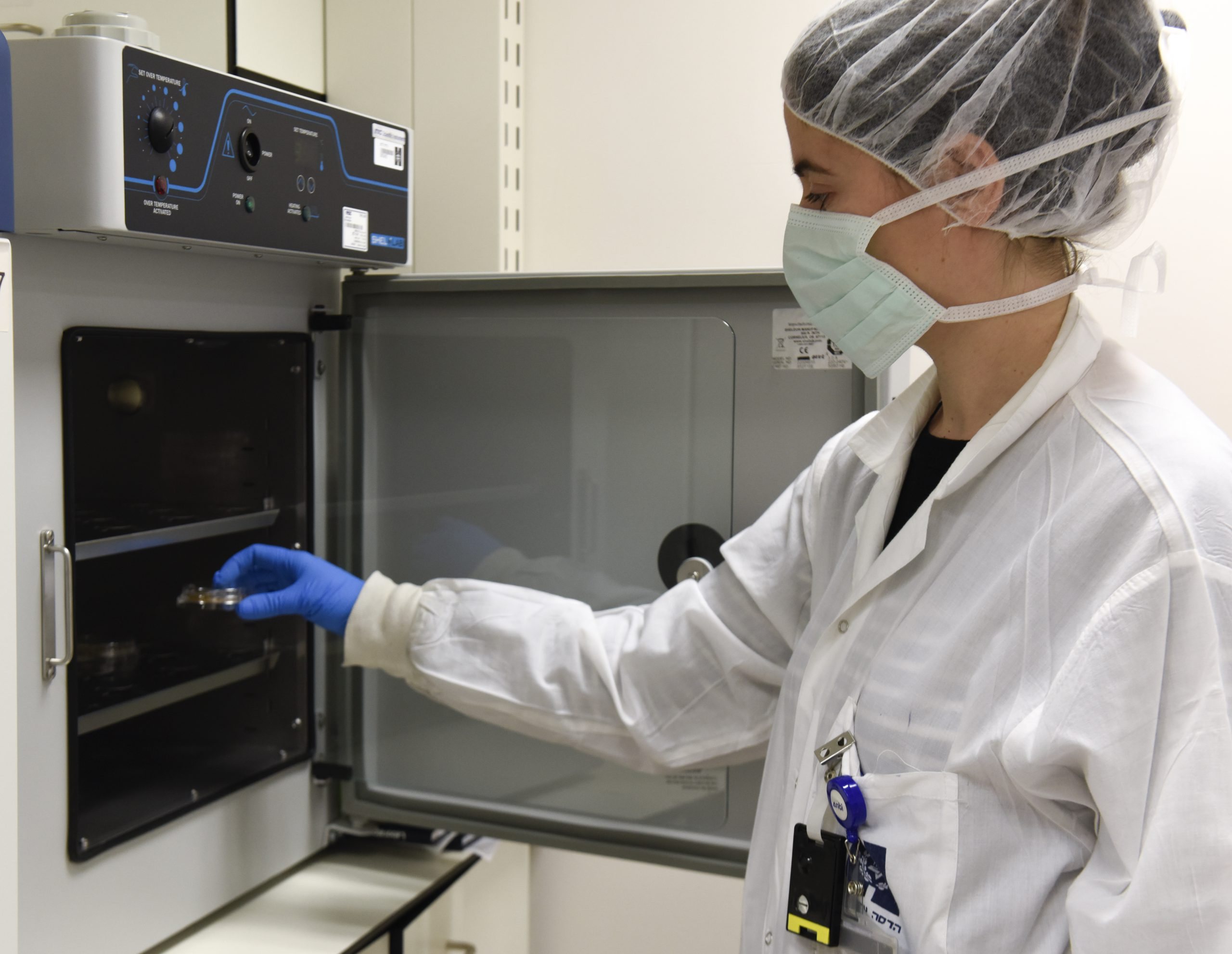
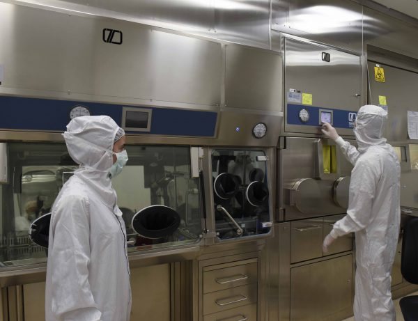
From here we reach the gleaming new production area, inaugurated just two months ago, meeting the strictest standards. Four years and about 13 million shekels were invested in building these rooms — and it shows. This is a whole other story; if until now we only put on shoe covers, here we’re covered head to toe, passing through doors that open before and close behind us. On the left, the “short-lived room,” on the right, the “long-lived room” – sounds like a joke about God or something, but it refers to the half-lives of radioactive materials.
Here, they separate the water, purify and collect the material, purify again, bottle it, pass it through a product chamber, put it in a small “lead fortress” that looks light as a Tiffin box but weighs over twenty kilograms. That gets put in an even bigger lead box, then sent out to customers: Assuta, Beilinson, Sheba, Ichilov, Shaare Zedek. Here you really feel the future, and the proof positive: no zip-ties here. Everything is impressive, all glass and lead.
After seeing how it’s made, we move to the nuclear medicine department- to see what’s done with it. Until now we’ve been in the realm of science, now we’re in medicine, with real people waiting after getting injected with the compounds we’ve just seen made. We’re brought to the new scanner, the first of its kind at Hadassah and in Israel — the digital PET-CT. Spectacular, this machine offers such high resolution that you can spot tumors as small as two or three millimeters – a 70% improvement in resolution, 90% in sensitivity. Also, you can inject the patient with less radioactive material, and they need to stay in the device for half the time, exposed to less than half the radiation.
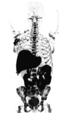
The arrival of the machine was accompanied by a minor scandal, when the Health Ministry had to step in to release it from customs, since it arrived before the installation permit was issued. But now it’s here and working. As I marvel at the new machine — there’s nothing like a new gadget – I notice there’s a patient inside. I don’t see his face, not exposed to his name, of course, but I see the imaging. It’s an incredible, truly three-dimensional view of the human body. Several black spots: brain, kidneys, bladder – where the glucose accumulates.
Then I see more and more black spots. These are metastases, cancerous growths spread throughout the body. Suddenly, it’s deeply depressing, thinking about the man in the machine whose face and name I don’t know. They show me more and more tumors of various types. Let me tell you, my friends, there are better ways to spend an hour. Eventually it gets quite grim, and we return to the surface.
Glossary | Guide
Isotope: A variant of an element, differing in neutron number and thus in mass. An isotope with too few neutrons is unstable and becomes radioactive.
Molecule: A structure built from several atoms connected by chemical bonds. Molecules usually have no electric charge; if they do, they’re called ions.
Neuroendocrine Tumors: Tumors that develop in neuroendocrine cells scattered throughout the body, and can produce hormones.
Curie (Ci): A unit of radioactivity named after Pierre and Marie Curie, discoverers of radium.
Nuclear Medicine: A field using radioactive atoms and ionizing radiation for research, imaging, diagnosis, and therapy. The radioactive atoms must emit enough radiation and have an appropriate half-life for imaging, yet without causing harm.
Half-life: Reflects an isotope’s stability; the time required for half of the atoms to decay. Can range betweeb fractions of a second to billions of years.
PET-CT: A test combining CT (using X-rays for anatomical results) and PET (which maps function via injection and tracking of a radioactive compound). Mostly used for diagnosing tumors.
Original article: Globes – Journey to the Belly of the Earth: The Fortified Particle Accelerator Fighting Cancer