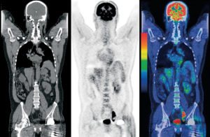
PET is an imaging technique in nuclear medicine that enables visualization of metabolic and molecular processes in the body and provides quantitative information about them. The scanner’s detectors identify pairs of photons with a unique energy, emitted simultaneously as a result of the radioactive decay of the administered radiopharmaceutical. At the end of the scan/imaging session, three-dimensional images are reconstructed, providing information on the distribution of the radiopharmaceutical in the body and its concentration in the target organ(s).
Currently, there are hybrid PET-CT and PET-MRI scanners that combine PET scan with CT or MRI, respectively. Not only does the CT/ MRI component provide crucial anatomical information, but it also enhances the quantification of PET data. Both CT / MRI and PET scans are performed on the same device. While the PET scan yields functional information (e.g., blood flow to an organ, its level of function, or the expression of a particular protein), the CT/ MRI scan provides anatomical information about the structure and location of organs, tumors, etc. Because both scans are performed using a single device, they deliver comprehensive information facilitating disease diagnosis, staging, and identification of unique molecular markers for various disease states.
For these reasons, and despite the relatively high costs of PET, this modality is commonly employed.